MRI
Magnetic resonance imaging (MRI), nuclear magnetic resonance imaging (NMRI), or magnetic resonance tomography (MRT) is a medical imaging technique used in radiology to investigate the anatomy and physiology of the body in both health and disease. MRI scanners use magnetic fields and radio waves to form images of the body. The technique is widely used in hospitals for medical diagnosis, staging of disease and follow-up without exposure to ionizing radiation
MEG/EEG
The electrical activity of active nerve cells in the brain produces currents spreading through the head. These currents also reach the scalp surface, and resulting voltage differences on the scalp can be recorded as the electroencephalogram (EEG). The currents inside the head produce magnetic fields which can be measured above the scalp surface as the magnetoencephalogram (MEG). EEG and MEG reflect brain electrical activity with millisecond temporal resolution, and are the most direct correlate of on-line brain processing obtainable non-invasively. Unfortunately, the spatial resolution of these methods is limited for principal physical reasons. Even with an infinite amount of EEG and MEG recordings around the head, a non-ambiguous localization of the activity inside the brain would not be possible. This “inverse problem” is comparable to reconstructing an object from its shadow: only some features (the shape) are uniquely determined, others have to be deduced on the ground of additional information. However, by imposing reasonable modeling constraints or by focusing on rough features of the activity distribution, useful inferences about the activity of interest can be made
PET
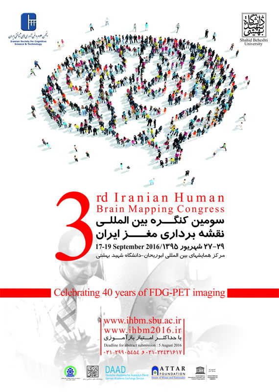
Positron emission tomography (PET) is a nuclear medicine, functional imaging technique that produces a three-dimensional image of functional processes in the body. The system detects pairs of gamma rays emitted indirectly by a positron-emitting radionuclide (tracer), which is introduced into the body on a biologically active molecule. Three-dimensional images of tracer concentration within the body are then constructed by computer analysis. If the biologically active molecule chosen for PET is fluorodeoxyglucose (FDG), an analogue of glucose, the concentrations of tracer imaged will indicate tissue metabolic activity as it corresponds to the regional glucose uptake. Use of this tracer to explore the possibility of cancer metastasis (i.e., spreading to other sites) is the most common type of PET scan in standard medical care (90% of current scans). However, on a minority basis, many other radioactive tracers are used in PET to image the tissue concentration of other types of molecules of interest. One of the disadvantages of PET scanners is their operating cost
Optic Imaging
Optical imaging is a technique for non-invasively looking inside the at the brain, which use ionizing radiation, optical imaging uses visible light and the special properties of photons to obtain detailed images of organs and tissues as well as smaller structures including cells and even molecules
Electrophysiology
Electrophysiology is the study of the electrical properties of biological cells and tissues. It involves measurements of voltage change or electric current on a wide variety of scales from single ion channel proteins to whole organs like the heart. In neuroscience, it includes measurements of the electrical activity of neurons, and particularly action potential activity. Recordings of large-scale electric signals from the nervous system such as electroencephalography may also be referred to as electrophysiological recordings
اطلاعات همایش برگرفته از وب سایت رسمی کنفرانس می باشد.
نویسنده: ص. م.
 اطلاع رسانی همایش و کنفرانس های علمی، معتبرترین همایش و کنفرانسهای دانشگاهها پایگاه خبری اطلاع رسانی همایش و کنگره و کنفرانس های کشور


